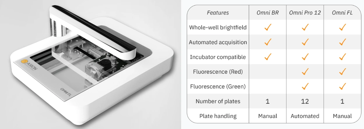
Description
Key Features
Cell Analysis in Brightfield and Fluorescence: The Omni facilitates cell analysis, offering dynamic visual results for a wide range of experiments, from label-free cell monitoring to fluorescence-based assays.
Continuous Monitoring from Your Incubator: Operating seamlessly within your incubator, the Omni automatically captures images as your cells thrive in their optimal conditions.
Comprehensive Cell Observation: The Omni’s innovative design moves the camera, not the cells, ensuring detailed brightfield images of the entire cell culture without disrupting cellular integrity.
Remote Cell Monitoring and Analysis: Its user-friendly software allows you to remotely monitor your cells and conduct data analysis right from your desktop, providing convenience and flexibility.
Quick and Easy Setup: The Omni is hassle-free with easy installation, zero maintenance requirements, and no calibration needed, making it accessible with just a brief training session.
Overview
The Omni platform is meticulously engineered to streamline and expedite the collection of intricate biological data. By harnessing the capabilities of brightfield and fluorescence imaging in conjunction with advanced software tools, the Omni empowers you to noninvasively and instantly scrutinize cell well-being and functionality in real-time.

Software/Algorithms
The Omni platform offers five distinct software modules tailored to cater to your specific assay requirements:
Cell Confluence: Available in both brightfield and fluorescence modes, this module equips you with the ability to precisely determine the optimal time for cell passaging or to monitor cell growth and viability in your experiments.
Wound Healing/Scratch Assay: Explore the dynamic progression of wound closure over time, gaining valuable insights into cellular migration and tissue repair processes.
Clonogenic Assay: This module enables you to effectively monitor and quantify cell proliferation and colony formation, facilitating the study of the impacts of cytotoxic and immunotherapeutic agents.
Fluorescent Object Count: Quantify fluorescent objects over time and across different wells while conducting fluorescence-based assays, ensuring accurate measurement of dynamic cellular responses.
Organoid Analysis: Utilizing an advanced machine-learning algorithm, this module empowers you to identify, track, and comprehensively analyse organoids across various culture vessels, opening up new possibilities for your research.
FAQ
How does the Omni work?
The Omni operates by illuminating samples using an LED and recording them with a moving camera positioned beneath the sample stage. During brightfield acquisition, the camera scans the sample stage, capturing a sequence of sequential images. A complete brightfield scan typically produces around 7850 snapshots, which are then stitched together to create an image with a surface area of 86 mm × 124 mm (3.4’’ × 4.9’’). When acquiring fluorescence images, users have the flexibility to choose how many snapshots of a specific location within the well to record. These images are subsequently uploaded to the Cloud, where they can be analyzed using our image analysis algorithms or compatible third-party software.
What image analysis modules are compatible with the Omni platform?
The Omni is compatible with a range of image analysis modules, including the Brightfield/Fluorescence Cell Confluence Analysis Algorithm, Scratch Assay (collective cell migration) Analysis Algorithm, Clonogenic Assay Algorithm, and Fluorescent Object Count. Additionally, users have the option to download raw data and conduct their own analysis using third-party software.
Is it possible to utilize the Omni platform within a cell culture incubator?
Certainly, Omni systems are purposefully designed for use within a cell culture incubator. The hardware and electronics are capable of operating within a temperature range of 5-40°C and under humidity conditions between 20-95%.
Which types of culture vessels can be used with the Omni systems?
The Omni systems can accommodate any transparent culture vessel with a height lower than 55 mm (2.2’’) (height of the light arc). This includes various containers such as 6- to 384-well plates, Petri dishes, and T25 to T225 flasks. It’s important to note that the scan area size is limited to 86 mm × 124 mm (3.4’’ × 4.9’’).


Reviews
There are no reviews yet.